Estimating liver perfusion parameters from triphasic CT
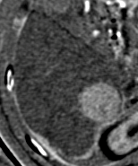
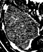
We developed a method for estimating the hepatic artery and portal vein blood supply to each portion of the liver, using standard triphasic CT. These perfusion parameters provide an estimate of the fraction of each pixel that is occupied by microscopic branches of the hepatic artery and portal vein. Using this method, we were able to quantitate the increase in hepatic artery perfusion in hepatocellular carcinoma (see figure), and the decrease in portal perfusion with cirrhosis.
| Color | Hepatic artery supply | Portal vein supply |
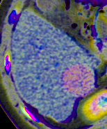 |  |  |
In the color image, the color indicates the phase of enhancement (arterial is red, portal venous is blue, and delayed is green). The saturation indicates the degree of enhancement (gray is non-enhancing, and color is enhancing). The brightness indicates the Hounsfield units.
Liver perfusion imaging detects:
- Early cirrhotic changes that are not reflected in Child Pugh score, and that predict survival.
- Aggressiveness of HCC that is not detected on core biopsy, and that predicts survival.
- Response to radioembolization of colorectal liver metastases.
View slides
Scroll through perfusion images on a web PACS
Perfusion calculator
References
Borgheresi A, Gonzalez-Aguirre A, Brown KT, Getrajdman GI, Erinjeri JP, Covey A, Yarmohammadi H, Ziv E, Sofocleous CT, Boas FE. (2018) "Does enhancement or perfusion on pre-procedure CT predict outcomes after embolization of hepatocellular carcinoma?" Academic Radiology. 25(12): 1588-94.
Boas FE, Brody LA, Erinjeri JP, Yarmohammadi H, Shady W, Kishore S, Sofocleous CT. (2016) "Quantitative enhancement measurements on preprocedure triphasic CT can predict response to radioembolization of colorectal liver metastases." AJR. 207: 671-5.
Boas FE, Kamaya A, Do B, Desser TS, Beaulieu CF, Vasanawala SS, Hwang GL, Sze DY. (2015) "Classification of hypervascular liver lesions based on hepatic artery and portal vein blood supply coefficients calculated from triphasic CT scans." Journal of Digital Imaging. 28: 213-23. [ Author's version ]
Return to the main page